On Tuesday May 9th the Integrated Imaging Facility held its 4th Annual Midwest Imaging and Microanalysis Workshop. During this workshop awards were presented for the 2016 best image publication. Two awards are given annually and each year the selection pool continues to grow more and more impressive. NDIIF is honored to be a part of the path to success that each of our investigators are on. It is our mission to continue to expand by developing new techniques and acquiring cutting-edge instrumentation to further the work and research of all of our users. The idea of including a “best artistic image award” has been considered for future award programs. We appreciate that many wonderful images have been acquired in our facilities but do not always make it into a publication for various reasons. However, these images are works of art and are an inspiration as well as an excellent showcase of the majestic features of science in motion and the power of our microscopes within NDIIF at the University of Notre Dame. Please join us in congratulating the 2016 winners and many thanks to all of those who submitted an image.
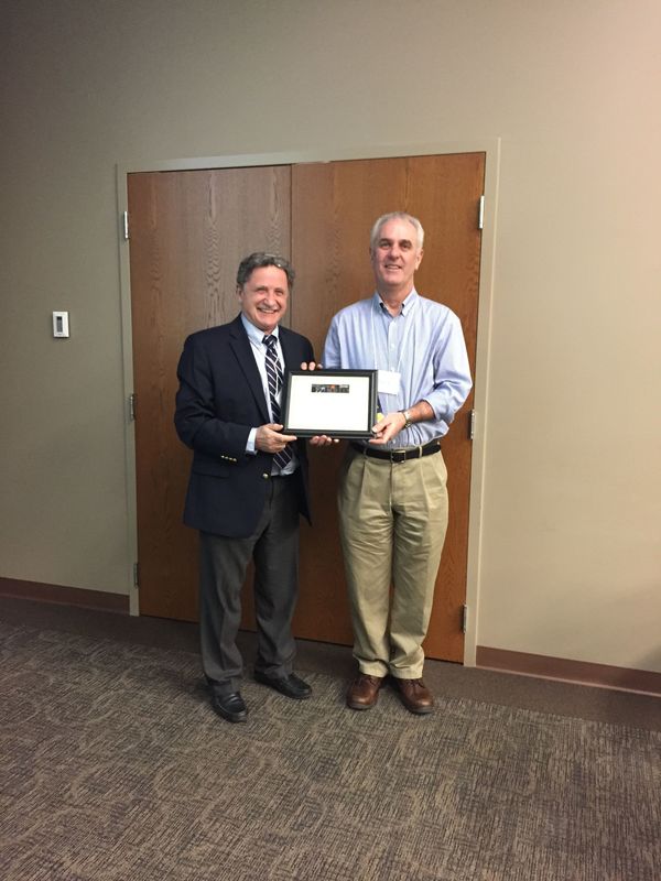
The Best Electron Beam Imaging Publication 2016 is awarded to Christopher Shuck, a graduate student working with Professor A. Mukasyan in the Department of Chemical and Biomolecular Engineering. Shuck and coworkers published a paper entitled “Ni/Al Energetic Nanocomposites and the Solid Flame Phenomenon”. The study work used a Focused Ion Beam (FIB) instrument to collect thousands of images of the nanocomposite Ni/Al system with nanometer accuracy. Using these images, the internal structure of the nanocomposite material was quantitatively analyzed using 3D reconstruction techniques. They determined surface contact between the reactants, layer thickness distributions, and then related them in a quantitative fashion to observed properties. The study was published in The Journal of Physical Chemistry C, 2016, 120, 27066.
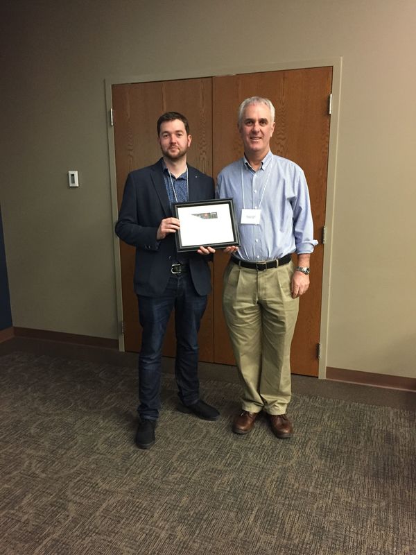
The Best Biological Imaging Publication 2016 is awarded to Dr. Eamonn Kennedy, apost-doctoral fellow collaborating with Professor G. Timp in the Departments of Electrical Engineering and Biological Sciences. Kennedy and coworkers published a paper entitled “Live Bacterial Physiology Visualized with 5 nm Resolution Using Scanning Transmission Electron Microscopy”. The study addressed a major limitation of transmission electron microscopy (TEM), that living cells cannot be maintained under the high vacuum imaging conditions. Combining live cell fluorescence microscopy with a new technique that permits TEM analysis within a sealed chamber of liquid held inside the Titan microscope, the team was able to visualize at nanometer resolution the infection of a living bacterial cell with bacteriophage virus without compromising cell viability. The study was published in ACS Nano, 2016, 10, 2669.E. Kennedy, Edward Nelson, T. Tanaka, J. Damiano, and G. Timp, "Live Bacterial Physiology Visualized with 5nm Resolution Using Scanning Transmission Electron Microscopy, ACS Nano 2016, Volume 10, pp. 2669-2677. DOI: 10.1021/acsnano.5b07697.
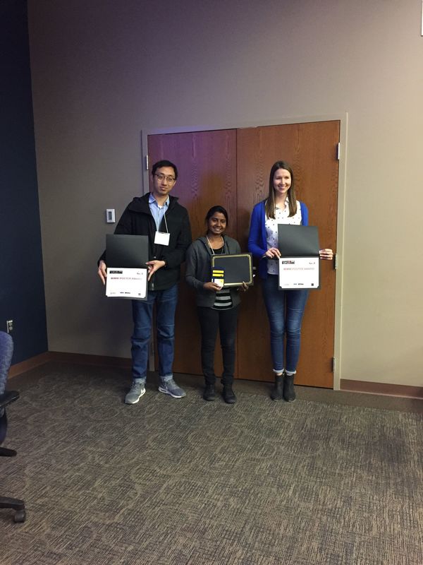
Also Best Poster Awards were presented at the Workshop! Biological Imaging posters as well as Electron Microscopy Posters were welcomed at the conference and the results were incredible. We appreciate the hard work and the dedication of our students and post-doctoral fellows. Please continue with your efforts and thank you for helping to make this workshop a wonderful success!
Originally published by at imaging.nd.edu on May 11, 2017.